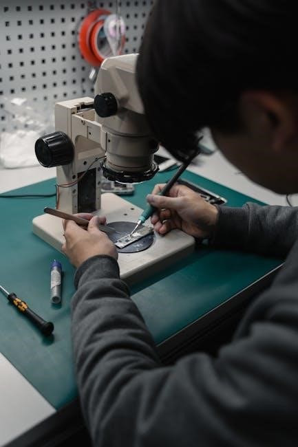
A microscope is an essential scientific tool designed to produce magnified images of small objects, capturing details invisible to the naked eye while minimizing distortion.
1.1 What is a Microscope?
A microscope is a scientific instrument designed to magnify small objects or samples, revealing details invisible to the naked eye. It works by focusing light through lenses to produce an enlarged image of the specimen. Microscopes are widely used in biology, medicine, and materials science to study microscopic structures. They consist of optical and mechanical components that work together to provide clear and precise images. The primary function of a microscope is to enhance visual observation, enabling scientists and researchers to analyze specimens in greater detail. By combining magnification and illumination, microscopes play a crucial role in advancing knowledge in various scientific fields.
1.2 Importance of Understanding Microscope Parts and Functions
Understanding the parts and functions of a microscope is essential for effective use and maintenance. Familiarity with components like the eyepiece, objective lenses, stage, and focus knobs ensures proper specimen preparation and observation. This knowledge helps users optimize image quality, prevent damage, and perform accurate adjustments. Additionally, understanding the microscope’s mechanics enhances efficiency in scientific workflows, enabling researchers to focus on analysis rather than operational challenges. Regular maintenance, such as cleaning optical parts and storing the microscope correctly, also relies on a solid understanding of its components. Thus, grasping the fundamentals of microscope anatomy and operation is crucial for both novice and experienced users to maximize its utility in scientific investigations and education.
Structural Parts of the Microscope
The microscope’s structural components include the base, arm, and stage with stage clips, providing stability and support for the instrument and its optical systems during operation.
2.1 Base of the Microscope
The base of the microscope serves as the foundation, providing stability and support for the entire instrument. It is typically made of heavy, durable materials to prevent vibrations and ensure steady operation. The base houses the light source, which illuminates the specimen, and connects to the arm and stage, holding them securely in place. Its robust construction is essential for maintaining the integrity of the microscope’s alignment and focus. Properly designed, the base ensures that the microscope remains stable during use, allowing for accurate observations and minimizing the risk of damage to the instrument or its components. Its importance lies in its role as the structural anchor of the microscope.
2.2 Arm of the Microscope
The arm of the microscope connects the base to the body tube, serving as a structural link between the stable foundation and the optical components. It is designed to be robust and rigid, ensuring minimal vibration and maintaining the alignment of the microscope’s parts. The arm supports the stage, focus knobs, and other essential components, allowing for smooth movement during specimen observation. Its durability and precise engineering are critical for maintaining the microscope’s stability and focus, enabling clear and accurate visualization of specimens. The arm’s design plays a vital role in the overall functionality of the microscope, ensuring that all parts work harmoniously to deliver optimal performance.
2.3 Stage and Stage Clips
The stage is a flat platform located below the microscope’s body tube, designed to hold specimens securely in place for observation. It is typically made of metal or durable materials to ensure stability. Stage clips, attached to the stage, grip the edges of microscope slides, preventing movement during examination. These clips are adjustable, allowing for precise alignment of the specimen under the objective lens. The stage’s stability is crucial for clear visualization, as any movement could distort the image. Additionally, the stage often features a mechanical stage control, enabling precise movement of the specimen along the X and Y axes, enhancing focusing accuracy and ease of use. Proper use of the stage and clips ensures optimal specimen positioning and secure handling.
Optical Components of the Microscope
The optical components, including the eyepiece, objective lenses, and revolving nosepiece, work together to magnify and clarify images, ensuring precise specimen visualization and detailed observation.
3.1 Eyepiece (Ocular Lens)
The eyepiece, or ocular lens, is a critical optical component positioned at the top of the microscope. Its primary function is to further magnify the image produced by the objective lens, enhancing the overall magnification power of the microscope. In binocular microscopes, two eyepieces are provided, allowing for comfortable viewing with both eyes. The eyepieces are typically interchangeable and come in various magnifications, such as 10x or 15x. They also feature a diopter adjustment, enabling users to fine-tune focus for their individual vision. Proper alignment and adjustment of the eyepiece ensure sharp, clear imagery, while regular cleaning and maintenance preserve its optical clarity and performance.
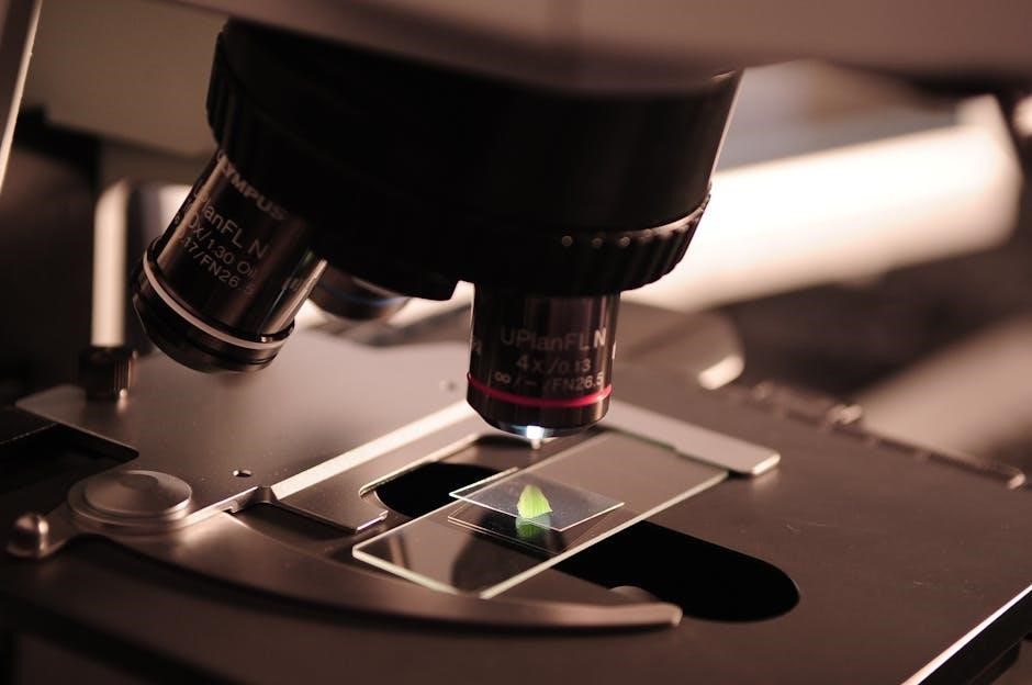
3.2 Objective Lenses
Objective lenses are a fundamental component of a microscope, responsible for collecting light from the specimen and forming an intermediate image. They are mounted on a revolving nosepiece, allowing easy switching between different magnifications. Common objective lenses include 4x, 10x, 40x, and 100x magnifications, with higher magnification lenses featuring longer barrels for increased working distance. The objective lens directly impacts the resolution and clarity of the image, making it essential for detailed observations. Proper alignment and maintenance of the objective lenses are critical to ensure optimal performance and prevent distortion. Regular cleaning and handling with care are recommended to preserve their optical quality and functionality over time.
3.3 Revolving Nosepiece
The revolving nosepiece is a circular, rotatable component that holds multiple objective lenses of varying magnifications. Its primary function is to allow seamless switching between different objective lenses without disturbing the specimen. By rotating the nosepiece, users can easily change the magnification power, ensuring efficient and precise observations. The nosepiece is typically designed with a click-stop mechanism to secure the objective lenses in position, preventing movement during use. This feature enhances stability and maintains focus, ensuring sharp and clear images. Proper alignment and maintenance of the revolving nosepiece are essential for optimal microscope performance and longevity of the optical components; Regular cleaning and lubrication of the mechanism are recommended to maintain smooth operation over time.
Focusing Mechanisms
Focusing mechanisms, including coarse and fine adjustment knobs, enable precise control over the microscope’s focus. They work together to achieve clear, sharp images of specimens at various magnifications.
4.1 Coarse Adjustment Knob
The coarse adjustment knob is a large, round control located on the side of the microscope. Its primary function is to move the stage up and down rapidly, allowing users to bring the specimen into general focus. This knob is typically used initially to approximate the focal plane, making it easier to locate the specimen under low magnification. Once the specimen is roughly in focus, the fine adjustment knob takes over for precise adjustments; Proper use of the coarse adjustment knob ensures that the specimen remains within the focal range, preventing damage to the objective lens or the slide. Regular maintenance, such as lubricating the mechanism, ensures smooth operation and extends the lifespan of the microscope. By mastering the use of the coarse adjustment knob, users can efficiently navigate the focusing process, enhancing their overall microscopy experience.
4.2 Fine Adjustment Knob
The fine adjustment knob is a smaller, precise control used to refine the focus of the specimen after the coarse adjustment knob has been used. Located alongside the coarse knob, it allows for minute adjustments to the stage’s position, ensuring the image is sharp and clear. This knob is essential for achieving high-resolution viewing, as it minimizes movement of the stage, preventing disruption of the specimen. The fine adjustment knob works in conjunction with the coarse knob, enabling users to transition smoothly from initial focusing to final precision. Proper use of this knob is critical for optimizing image clarity and maintaining the integrity of the microscope’s optical system. Regular cleaning and lubrication of the fine adjustment mechanism are recommended to ensure optimal performance and longevity.
Illumination and Light Control
Illumination systems in microscopes include a light source, condenser, and diaphragm. These components work together to focus and regulate light, optimizing specimen visibility under magnification.
5.1 Light Source (Lamp)
The light source, often a built-in lamp, is crucial for illuminating specimens. It provides sufficient light to pass through the sample, enhancing visibility. Positioned near the base, it ensures even light distribution. Proper adjustment of the light intensity is essential for clear imaging. Modern microscopes may use LED lamps for brighter, energy-efficient illumination. The light source works in conjunction with the condenser and diaphragm to optimize light quality and focus. Without adequate lighting, detailed observation becomes challenging, making the light source a fundamental component for effective microscopy.
5.2 Condenser and Diaphragm
The condenser focuses light onto the specimen, ensuring even illumination. Positioned below the stage, it can be adjusted vertically to optimize light intensity. The diaphragm, a rotating disk with varying aperture sizes, regulates the amount of light reaching the specimen. By adjusting the diaphragm, users can control light intensity, reduce glare, and enhance contrast for clearer observations. Together, the condenser and diaphragm are essential for achieving proper lighting conditions, which are critical for high-quality microscopy. Their precise alignment and adjustment ensure optimal light transmission, making them indispensable for detailed specimen analysis.

Additional Features of Modern Microscopes
Modern microscopes include advanced features like diopter adjustment for individual eye calibration and field aperture control to optimize light entry, enhancing visibility and adaptability for diverse specimens.
6.1 Diopter Adjustment
Diopter adjustment is a feature in binocular microscopes that allows users to fine-tune the focus for each eyepiece, accommodating differences in vision between the two eyes. This ensures a clear, single image when viewing specimens. By adjusting the diopter, individuals with varying refractive errors can achieve a sharp focus without relying on corrective eyewear. This feature enhances comfort and precision during extended observation. Proper use of the diopter adjustment involves setting the eyepieces to match the user’s specific visual needs, ensuring optimal performance and reducing eye strain during microscopy. This customization is particularly useful in educational and professional settings, where microscopes are shared among multiple users with diverse visual requirements.
6.2 Field Aperture
The field aperture, located in the condenser, regulates the amount of light entering the microscope. It adjusts the diameter of the light beam, optimizing illumination for the specimen. Proper adjustment ensures even light distribution, enhancing image clarity and contrast. The field aperture works in conjunction with the condenser to focus light precisely on the sample, minimizing glare and improving visibility. Correctly setting the field aperture is critical for achieving sharp, well-illuminated images, especially when using higher magnification objectives. This feature is essential for maintaining optimal viewing conditions and ensuring accurate observations during microscopy.

Mechanical Components
Mechanical components include the body tube, which holds optical parts, and the focus lock, stabilizing the arm. The XYZ plane function enables precise movement for accurate focusing.
7.1 Body Tube
The body tube is a critical mechanical component of the microscope, housing the optical system. It connects the eyepiece to the objective lenses, ensuring proper alignment and light transmission. The tube’s design supports the structural integrity of the microscope, maintaining the positions of the eyepiece and objective lenses. This alignment is essential for clear and focused imaging. The body tube also plays a role in the overall durability of the microscope, protecting internal components from external damage. Proper maintenance of the body tube is vital to preserve the microscope’s functionality and optical performance over time. Its stability ensures consistent and precise observations during use.
7.2 Focus Lock and XYZ Plane Function
The focus lock and XYZ plane function are advanced features designed to enhance precision and control during microscopy. The focus lock ensures the microscope’s arm remains stable, preventing unintended movement that could disrupt focus. This is particularly useful during high-magnification observations. The XYZ plane function allows for precise adjustments in three dimensions, enabling accurate positioning of the specimen. Together, these features improve focus stability and specimen alignment, reducing the risk of damage to the microscope or the sample. These mechanisms are essential for maintaining clarity and consistency in microscopic examinations, especially in demanding applications such as biological research or materials science.

Operational Steps for Using a Microscope
Prepare the microscope, place the slide on the stage, adjust focus using coarse and fine knobs, and regulate illumination for clear specimen observation and analysis.
8.1 Preparing the Microscope for Use
Preparing the microscope ensures optimal performance and prevents damage. Begin by placing the microscope on a stable, flat surface. Clean all optical parts, including the eyepiece and objective lenses, using soft, lint-free cloth and appropriate cleaning solutions. Next, ensure the stage is clear and the stage clips are secure. Adjust the condenser to align with the objective lens and position it close to the stage. Turn on the light source and set the light intensity using the diaphragm. Finally, ensure the coarse adjustment knob is in the correct position for focusing. Proper preparation ensures clear visibility and accurate specimen observation.
8.2 Focusing on a Specimen
Focusing on a specimen involves a systematic approach to achieve clear observation. Begin by placing the prepared slide on the stage, securing it with stage clips. Using the coarse adjustment knob, lower the objective lens toward the slide until the specimen comes into view. Once the specimen is visible under low magnification, switch to a higher power objective lens for greater detail. Fine-tune the focus using the fine adjustment knob for clarity. Ensure the specimen is centered and evenly illuminated by adjusting the condenser and diaphragm. Proper focusing enhances image quality and ensures accurate observation of the specimen.
Maintenance and Care of the Microscope
Regular cleaning of optical parts with lens tissue prevents dust buildup. Store the microscope in a dry, secure location to protect its components and maintain functionality.
9.1 Cleaning the Optical Parts
Cleaning the optical parts of a microscope is crucial for maintaining clarity and performance. Use soft, dry optical lens tissue to gently wipe the eyepiece, objective lenses, and other glass surfaces. Avoid using chemicals, household cleaners, or abrasive materials, as they can damage the coatings or scratch the lenses. For stubborn smudges, lightly dampen the tissue with distilled water or a recommended cleaning solution. Never touch the lens surfaces with bare hands, as oils from skin can leave residue. Regular cleaning prevents dust buildup and ensures sharp, distortion-free images. Proper care extends the lifespan of the microscope and maintains its accuracy for precise observations.
9.2 Storing the Microscope Properly
Proper storage is essential to maintain the microscope’s performance and longevity. Always store the microscope in a cool, dry place, away from direct sunlight and moisture. Use a dust cover to protect the optical and mechanical components from dust and debris. Ensure the microscope is placed upright to prevent damage to the optics or electrical systems. Clean the microscope thoroughly before storage to prevent dust buildup. Store it in a secure, stable location to avoid accidental knocks or vibrations. For extended storage, consider using silica gel packets to maintain a dry environment. Proper storage ensures the microscope remains in optimal condition for future use.
A microscope is a powerful tool with intricate parts designed for magnification, light control, and focus. Understanding its components ensures effective use and proper maintenance for lasting functionality.
10.1 Summary of Key Components and Functions
The microscope is a sophisticated instrument with essential components working together to enhance observation. Key parts include the eyepiece, objective lenses, stage, and adjustment knobs. The eyepiece magnifies images, while objective lenses provide varying magnification levels. The stage holds specimens securely, and coarse and fine adjustment knobs focus the image. Additional components like the light source, condenser, and diaphragm control illumination. Understanding these parts and their functions ensures proper use, maintenance, and optimal performance of the microscope. Each component plays a vital role in delivering clear, detailed, and accurate observations, making the microscope an indispensable tool in scientific and educational settings.